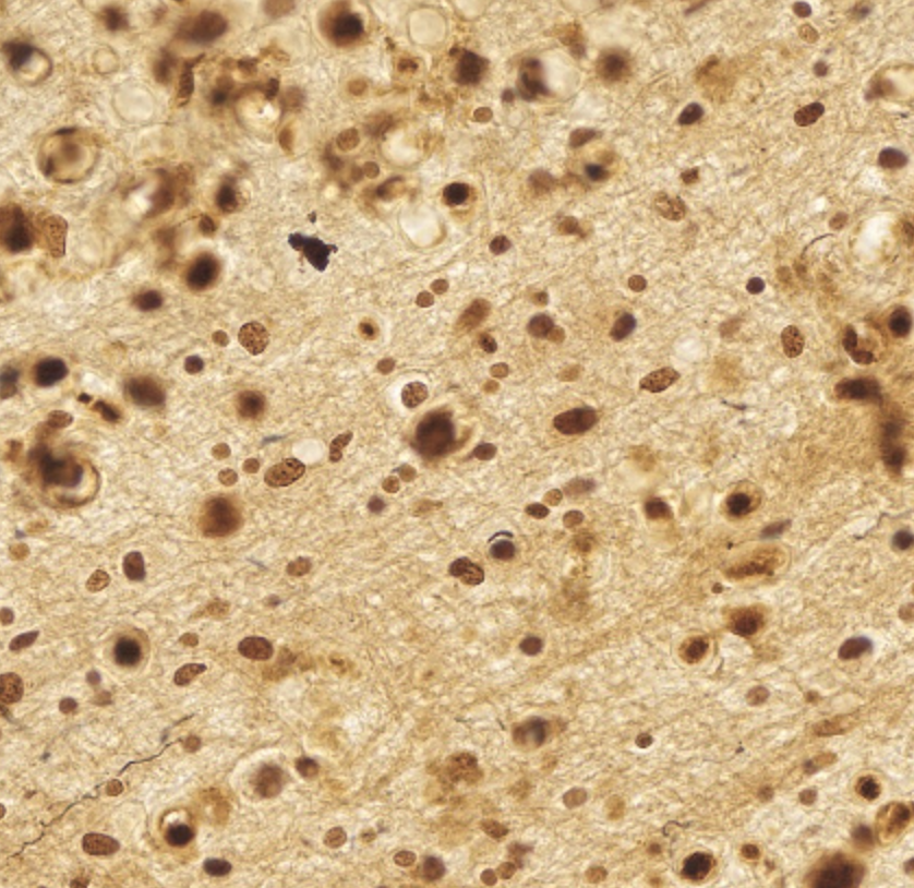
- Figure 1
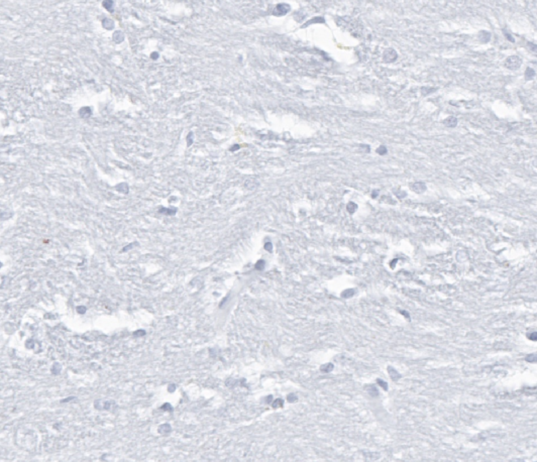
- Figure 2
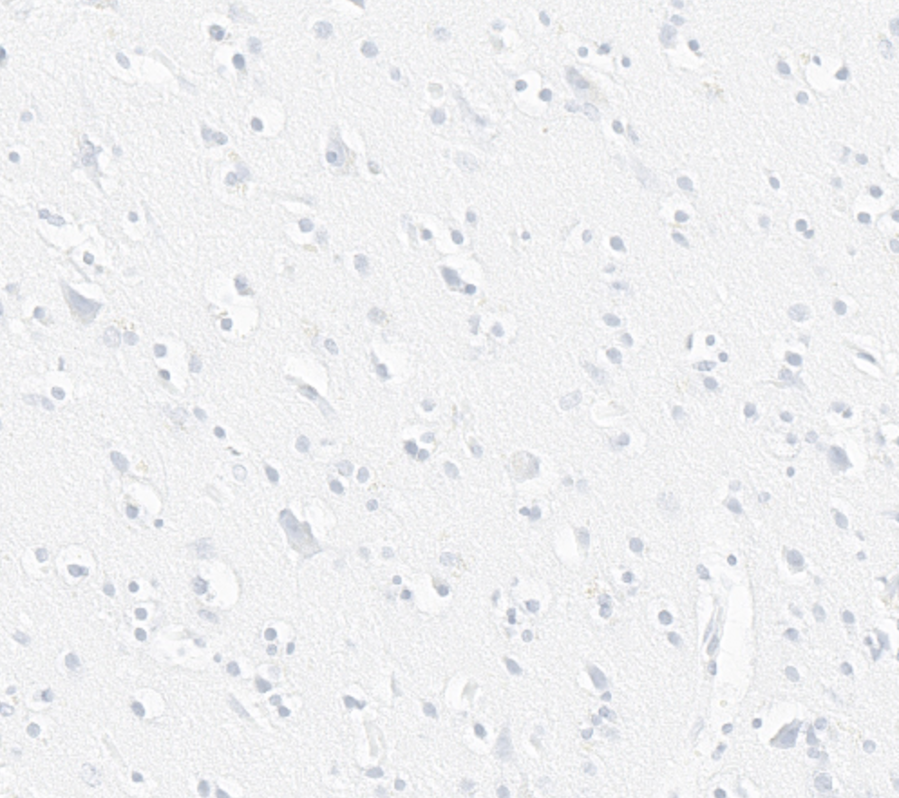
- Figure 3
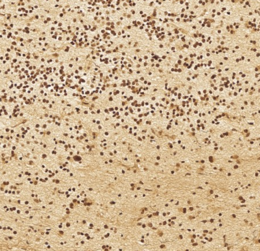
- Figure 4


Figure 1-4. These images are of parts of brain tissue taken from FHS participants who volunteered to donate their brains for research purposes. The neuropath data in the curated dataset comes from the same brains, where neuropathological evaluation of autopsied brains was performed and gross neuropathological findings were recorded. The full images are large .svs files, which are created by digital microscope scanners as zoomable digital images. (Further information coming soon)